Patient Education

What is an X-Ray Exam?
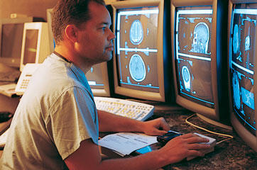
X-rays are forms of radiant energy, like light or radio waves. Unlike light, x-rays can penetrate the body, which allows a radiologist to produce pictures of internal structures. The radiologist can view these on photographic film or on a TV or computer monitor.
X-ray examinations provide valuable information about your health and play an important role in helping your doctor make an accurate diagnosis. In some cases x-rays are used to assist with the placement of tubes or other devices in the body or with other therapeutic procedures.
Measuring radiation dosage
The scientific unit of measurement for radiation dose, commonly referred to as effective dose, is the millisievert (mSv). Other radiation dose measurement units include rad, rem, roentgen, sievert, and gray.
Because different tissues and organs have varying sensitivity to radiation exposure, the actual radiation risk to different parts of the body from an x-ray procedure varies. The term effective dose is used when referring to the radiation risk averaged over the entire body.
The effective dose accounts for the relative sensitivities of the different tissues exposed. More importantly, it allows for quantification of risk and comparison to more familiar sources of exposure that range from natural background radiation to radiographic medical procedures.

Are X-Rays safe?
As with other medical procedures, x-rays are safe when used with care. Radiologists and x-ray technologists have been trained to use the minimum amount of radiation necessary to obtain the needed results. Properly conducted imaging carries minimal risks and should be performed when clinically indicated. The amount of radiation used in most examinations is very small and the benefits greatly outweigh the risk of harm.
X-rays are produced only when a switch is momentarily turned on. As with visible light, no radiation remains after the switch is turned off.
X-rays over your lifetime
The decision to have an x-ray exam is a medical one, based on the likelihood of benefit from the exam and the potential risk from radiation. For low dose examinations, usually those that involve only films taken by a technologist, this is generally an easy decision. For higher dose exams such as computed tomography (CT) scans and those involving the use of contrast materials (dyes) such as barium or iodine, the radiologist may want to consider your past history of exposure to x-rays. If you have had frequent x-ray exams and change healthcare providers, it is a good idea to keep a record of your x-ray history for yourself. This can help your doctor make an informed decision. It is also very important to tell your doctor if you are pregnant before having an exam that involves the abdomen or pelvic region.
Recently there have been a number of studies* linking current or future cancers to previous x-ray imaging studies, especially CT scans. These studies have important limitations in that they lack key data, including: direct radiation exposure measurements for each patient; why the patient underwent the study; and what beneficial information was derived from the CT scan. In addition, underlying statistical models can be fraught with tremendous levels of uncertainty. Nevertheless, these studies are valuable as they highlight the importance of optimizing CT scan techniques and have led to advancements that are resulting in much lower radiation exposures for similar CT studies.
Magnetic Resonance Imaging (MRI) During Pregnancy
If you are pregnant and your doctor wants to perform a magnetic resonance imaging (MRI) exam, there is a possibility that your doctor is concerned about your health or the health of your baby.
Illness during pregnancy
In general, doctors try to delay treating a medical problem, if possible, until after pregnancy. Therefore, if your doctor orders an MRI during your pregnancy, he/she is trying to address a potentially urgent or serious illness.
What to expect during the MRI exam?
Your exam will focus on imaging a particular body part, such as the baby in the pelvis, the brain or the spine. Scanning time varies depending on the body part being imaged; however, most exams will require that you lie still for 20 to 40 minutes.